The author: Professor Yasser Metwally
INTRODUCTION
October 11, 2008 — In this edition of the monthly publication “Topic of the month” Professor Metwally discusses the neuroimaging findings in pituitary adenomas. This topic is presented in downloadable PDF format.
Click here to download this monthly topic (Neuroimaging of pituitary adenomas) in PDF format (570 KB)
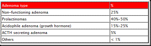
Originally termed chromophobe adenomas, endocrine-inactive pituitary tumors were once considered the largest group of pituitary tumors. With advances in endocrinologic testing and modern immunohistochemical and immunoelectron microscope techniques, the incidence of adenomas with no evidence of hypersecretion or endocrine activity has decreased to about 25 per cent of pituitary adenomas. Histologically, these adenomas have secretory granules and immunocytochemically are growth hormone or prolactin-positive, despite no associated clinical changes or abnormal serum hormone levels about 5 per cent of the time. Inactive tumors have cells with no histologic, immunocytologic, or electron microscopic markers (Null cells). They are chromophobic and electron microscopy show them to have poorly developed cytoplasm, indented nuclei, and sparse granules (100 to 250 lim) lined up along the cell membrane.
It is the functionally active group of pituitary tumors that comprise the largest percentage of pituitary adenomas. They represent about 75 per cent of all pituitary tumors. Preoperative endocrinologic testing, as well as clinical symptomatology resulting from the adenoma’s hypersecretion of hormones, helps to identify and classify these tumors. It is this functional classification confirmed with immunohistochemical and immunoelectromicroscopic techniques and not traditional light microscopic pathology that separates these tumors.
Prolactinomas represent about 40 to 50 per cent of all patients with pituitary adenomas. Under light microscopy, prolactin cell tumors are chromophobic or acidophilic. Using immunoelectron microscopy, they may be classified as densely or sparsely granular, although the former type is quite rare. The densely granular resemble nontumor lactotrophic pituitary cells that are resting and nonsecreting. The sparsely granular type resemble the nontumor lactotrophic pituitary cells that are actively secreting. Their secretary granules are sparse, spherical, and measure 150 to 350 nm.
Somototrophic adenomas, resulting in acromegaly, account for 15 to 25 per cent of pituitary adenomas. Under light microscopy, these tumors may be termed acidophilic or chromophobic. Using immunoelectron microscopy, two distinct cell types can be identified: densely and sparsely granulated adenomas. The densely granulated cell type more closely resembles nontumor pituitary somototrophic cells and is characterized by well-developed endoplasmic reticulum, permanent Golgi complexes, and numerous spherical densely staining secretary granules. The sparsely granulated type differ from nontumorous pituitary somototrophic cells in that it has permanent Golgi complexes, irregular nuclei, few spherical secretary granules, and several centrioles.
Cushing’s disease or Nelson’s syndrome caused by corticotropin-secreting adenomas represent only about 5 per cent of all pituitary adenomas. Under light microscopy, corticotrophs are basophilic. Immunoelectron microscopy shows these tumor cells to be similar to corticotrophic nontumorous pituitary cell types containing numerous spherical secreting granules that vary in density, measure 250 to 700 nm, and line up along the cell membranes.
The rarest of pituitary adenomas are those that secrete solely thryotrophin or gonadotropin. Each type accounts for less than 1 per cent of pituitary adenomas. Under light microscopy, the thyrotropic adenomas are chromophobic and under electron microscopy, they have long cytoplasmic processes, sparse, spherical secreting granules (150 to 250 nm), and abundant endoplasmic reticulum.
Pituitary macroadenomas are, by definition, at least 10 mm in size or more, while microadenomas are less than 10 mm in size.
Click here to download this monthly topic (Neuroimaging of pituitary adenomas) in PDF format (570 KB)
References
Metwally, MYM: Textbook of neuroimaging, A CD-ROM publication, (Metwally, MYM editor) WEB-CD agency for electronic publication, version 9.4a October 2008 [Click to have a look at the home page]

No comments:
Post a Comment